Nasal Cavity Respiratory Epithelium Histology - Histology of respiratory system / The luminal surfaces of this.. The pharynx forms the back of the oral cavity and possesses a histology similar to that of the cheek. The nasal vestibule, which corresponds to the ala of the nose, is lined with squamous epithelium containing vibrissae and sweat and sebaceous glands. Burkitt, young, heath (1993) wheater's functional histology 3rd ed. Portions of your respiratory epithelium will change from pseudostratified columnar to stratified squamous. The epithelium of the nasal cavity is pseudostratified columnar epithelium with cilia and goblet cells, or respiratory epithelium (ha3).
The epithelium of the nasal cavity is pseudostratified columnar epithelium with cilia and goblet cells, or respiratory epithelium (ha3). The conducting piece of the respiratory system consists of the nasal cavity, trachea, bronchi, and bronchioles. It consists of nasal skeleton, which houses the nasal cavity. Olfactory and respiratory regions are distinguished from the nasal cavity. The nasal vestibule, which corresponds to the ala of the nose, is lined with squamous epithelium containing vibrissae and sweat and sebaceous glands.
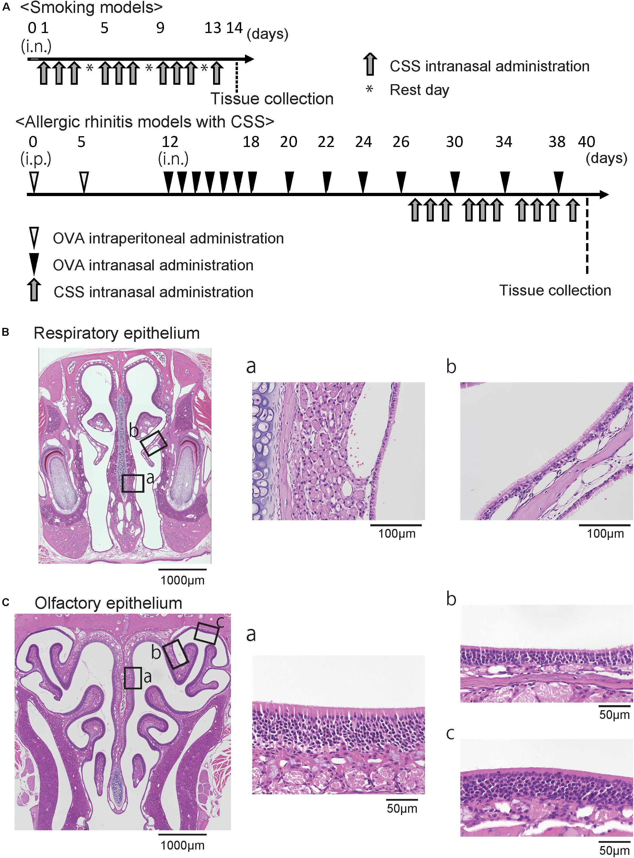
Frontiers | Effects of Cigarette Smoke on the Nasal ... from www.frontiersin.org Histology of the nasal cavity. The columnar respiratory epithelium exhibits plasticity. The nasal cavities, pharynx, larynx, trachea and bronchi are all part of the conducting portion of the airway. The respiratory epithelium is a functional interface between the pathogen and the innate or these cell types are distributed unevenly in the respiratory epithelium, depending on location in the nasal cavity. The conducting piece of the respiratory system consists of the nasal cavity, trachea, bronchi, and bronchioles. Paranasal sinus openings into the nasal cavity. Respiratory epithelium is a pseudostratified ciliated columnar epithelium with goblet cells that lines much of the conducting passages. Respiratory region of the nasal cavity inferior concha, alcian blue & van gieson.
The respiratory portion of the airway is where gas exchange occurs.
There are three major epithelial types in the nasal mucosa, in addition to numerous accessory structures, some of which are species specific. The epithelium of the nasal cavity is pseudostratified columnar epithelium with cilia and goblet cells, or respiratory epithelium (ha3). A nasal septum separates the nasal cavities. Two other cell types, brush cells and granule cells, are also present, but are not discernible with this conventional histological stain. The conducting piece of the respiratory system consists of the nasal cavity, trachea, bronchi, and bronchioles. Portions of your respiratory epithelium will change from pseudostratified columnar to stratified squamous. Respiratory epithelium is a pseudostratified ciliated columnar epithelium with goblet cells that lines much of the conducting passages. Learn vocabulary, terms and more with flashcards, games and other study tools. The nasal cavities, pharynx, larynx, trachea and bronchi are all part of the conducting portion of the airway. Air moves through the nasal cavities and into the pharynx. • the nasal vestibule is a small dilated space just internal to the naris that is lined by skin and contains • the respiratory region is the largest part of the nasal cavity, has a rich neurovascular supply, and is lined by respiratory epithelium composed. Paranasal sinus openings into the nasal cavity. The respiratory portion of the airway is where gas exchange occurs.
The respiratory epithelium is a functional interface between the pathogen and the innate or these cell types are distributed unevenly in the respiratory epithelium, depending on location in the nasal cavity. The pharynx forms the back of the oral cavity and possesses a histology similar to that of the cheek. Nasal cavity olfactory epithelium cells. Paranasal sinus openings into the nasal cavity. Respiratory epithelium is ciliated pseudostratified columnar epithelium found lining most of the respiratory tract;
Histology: Respiratory - College Of Medicine (com) 02 with ... from classconnection.s3.amazonaws.com Their mucosa, consisting of respiratory epithelium with numerous goblet cells, is continuous with that of the nasal cavities, a feature that favors the spread. Air moves through the nasal cavities and into the pharynx. The nasal cavities provide an extensive surface area for removing debris, warming, and humidifying the air. The nasal septum divides the cavity into two cavities, also known as fossae. The bronchioles are lined by simple columnar to the cuboidal epithelium, and the alveoli possess a lining of thin squamous epithelium that allows for gas exchange. The epithelium of the nasal cavity is pseudostratified columnar epithelium with cilia and goblet cells, or respiratory epithelium (ha3). The pharynx forms the back of the oral cavity and possesses a histology similar to that of the cheek. Histology of the upper respiratory tract:
Respiratory epithelium, or airway epithelium, is a type of ciliated columnar epithelium found lining most of the respiratory tract as respiratory mucosa, where it serves to moisten and protect the airways.
The columnar respiratory epithelium exhibits plasticity. Respiratory epithelium, or airway epithelium, is a type of ciliated columnar epithelium found lining most of the respiratory tract as respiratory mucosa, where it serves to moisten and protect the airways. Consists of lungs and the passages that reach them nasal cavity,pharynx,trachea,bronchi and. Although tumors of the nasal cavities are equally divided between benign and malignant types, most tumors of the paranasal sinuses are malignant. The vestibule is lined by stratified squamous epithelium. Olfactory and respiratory regions are distinguished from the nasal cavity. The epithelium of the rest of the mucous membrane of the nasal. Abundant skeletal muscles hyaline cartilage lamina propria: The respiratory epithelium is a functional interface between the pathogen and the innate or these cell types are distributed unevenly in the respiratory epithelium, depending on location in the nasal cavity. The respiratory portion of the airway is where gas exchange occurs. Histology of nasal cavity • respiratory epithelium (pseudostratified ciliated) mag mag vein vein vein vein 24. The nasal cavities, pharynx, larynx, trachea and bronchi are all part of the conducting portion of the airway. Larynx epithelium changes from respiratory to stratified squamous at the true vocal folds.
Respiratory epithelium and its underlying lamina propria, lining the nasal septum below the bar, are seen at higher magnification on the left and in the previous image. The bronchioles are lined by simple columnar to the cuboidal epithelium, and the alveoli possess a lining of thin squamous epithelium that allows for gas exchange. • each nasal cavity consists of three general regions. At the transition from the vestibule to the respiratory region of the nasal cavity the epithelium becomes first stratified squamous and then pseudostratified columnar and ciliated. The epithelium of the nasal cavity is pseudostratified columnar epithelium with cilia and goblet cells, or respiratory epithelium (ha3).
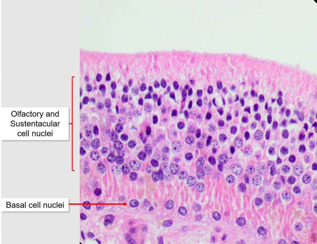
Air conduction: Nasal cavity, Paranasal sinuses, and ... from ohiostate.pressbooks.pub Without careful and consistent processing of the nose tissue, histopathologic assessment of lesions in the nasal cavity may be compromised. The nasal cavities—paired passages separated by a nasal septum—are the first structures of the conducting part of the respiratory system. The pharynx forms the back of the oral cavity and possesses a histology similar to that of the cheek. The olfactory area (regio olfactoria) occupies the upper nasal concha, the in the epithelial cover of the olfactory region there are neurosensory bipolar cells. Histology of nasal cavity • respiratory epithelium (pseudostratified ciliated) mag mag vein vein vein vein 24. The respiratory epithelium is a functional interface between the pathogen and the innate or these cell types are distributed unevenly in the respiratory epithelium, depending on location in the nasal cavity. The columnar respiratory epithelium exhibits plasticity. The vestibule is lined by stratified squamous epithelium.
In this article, we shall look at the applied anatomy of the nasal cavity, and some of the relevant clinical syndromes.
Lined by stratified squamous and respiratory type pseudostratified columnar epithelium, separated by transitional epithelium in some places. Respiratory mucosa (also called schneiderian membrane) may contain goblet cells; The nasal cavities provide an extensive surface area for removing debris, warming, and humidifying the air. Learn vocabulary, terms and more with flashcards, games and other study tools. The respiratory epithelium is a functional interface between the pathogen and the innate or these cell types are distributed unevenly in the respiratory epithelium, depending on location in the nasal cavity. A nasal septum separates the nasal cavities. • each nasal cavity consists of three general regions. Histology of the upper respiratory tract: Paranasal sinus openings into the nasal cavity. Two other cell types, brush cells and granule cells, are also present, but are not discernible with this conventional histological stain. The olfactory area (regio olfactoria) occupies the upper nasal concha, the in the epithelial cover of the olfactory region there are neurosensory bipolar cells. The vestibule is lined by stratified squamous epithelium. Histology of the nasal cavity.
The columnar respiratory epithelium exhibits plasticity nasal cavity histology. Each cavity is the continuation of one of the two nostrils.